
We accurately determine molecular weight and assess the structural integrity of your protein, by identifying potential modifications, degradation, and truncations.
Using UPLC-MS/MS, we perform detailed analysis of proteolytic digests to precisely locate and quantify PTMs, confirm sequence integrity, and support batch-to-batch comparability studies for your protein.
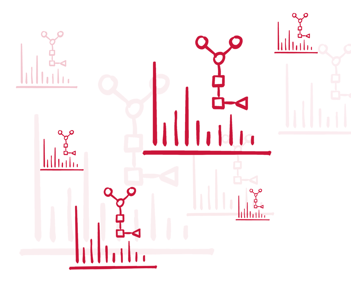
We provide structural insights at the subunit level through the characterization of antibody fragments (Fab/Fc), heavy and light chains, and associated glycoforms—ideal for complex molecules and biosimilarity assessments.
We determine the drug-to-antibody ratio (DAR), assess conjugation sites, and evaluate the distribution and stability of your ADC product.
We verify correct disulfide bond formation and identify scrambled or incorrect linkages, ensuring proper folding and structural stability of your protein, while considering plausible modification of the affected (redox-sensitive) cysteines.

